

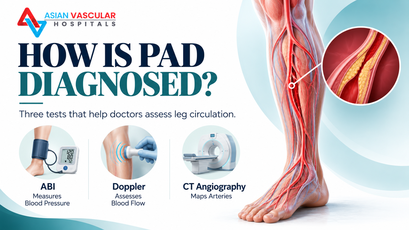
How Is Peripheral Artery Disease Diagnosed?
Peripheral artery disease (PAD) occurs when arteries supplying blood to the legs become narrowed or blocked, most commonly because of atherosclerosis. It can cause leg pain while walking, reduced walking distance, cold feet, slow-healing wounds and, in advanced cases, pain at rest.
PAD diagnosis tests help doctors determine whether blood flow is reduced, identify the location and severity of arterial disease, and decide whether further treatment or imaging is needed.
The evaluation commonly begins with a medical history, physical examination and an ankle-brachial index (ABI) test. Depending on the findings, a doctor may recommend Doppler or duplex ultrasound, CT angiography (CTA), or other specialised tests.
Not every patient needs every test. The appropriate investigation depends on symptoms, examination findings, medical history and whether an intervention is being considered.
If you are experiencing leg pain while walking or other possible circulation symptoms, read our guide to peripheral artery disease symptoms and early warning signs.
What Are the Main Tests for Peripheral Artery Disease?
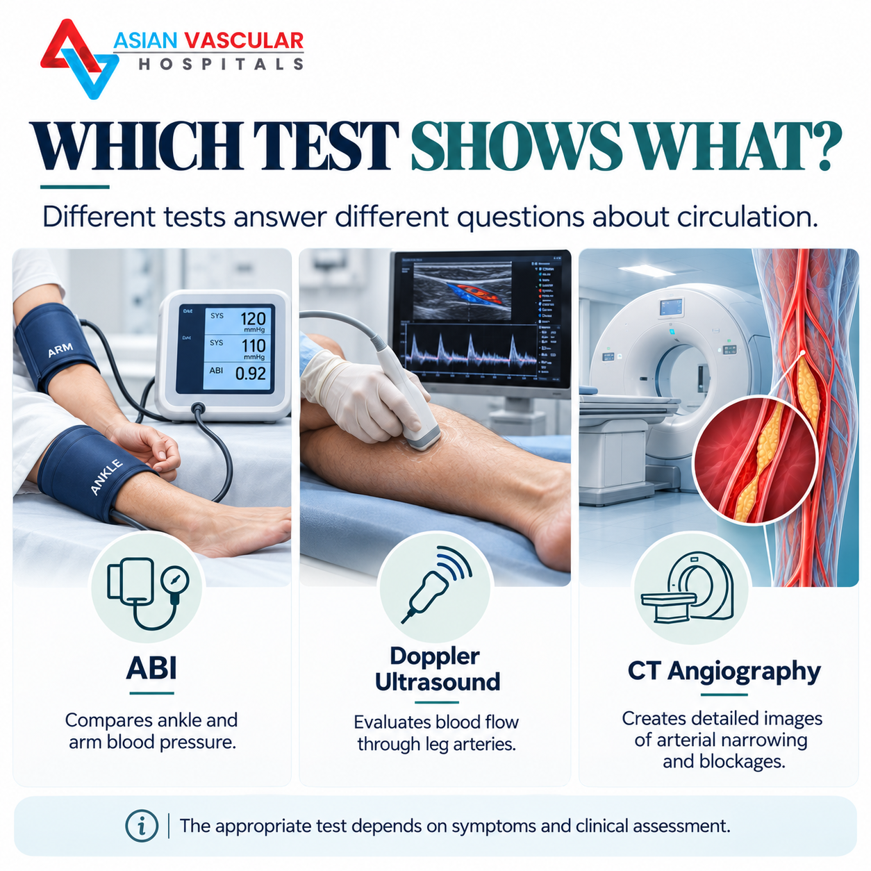
Doctors use different investigations to answer different questions. Some measure how well blood reaches the legs, while others create images of the arteries.
| Test | What it evaluates | When it may be used |
|---|---|---|
| Ankle-brachial index (ABI) | Compares blood pressure at the ankle and arm | Common initial test when PAD is suspected |
| Doppler or duplex ultrasound | Assesses blood flow and can identify narrowed or blocked arterial segments | When the location and flow characteristics of disease need evaluation |
| CT angiography (CTA) | Produces detailed images of blood vessels | When detailed arterial mapping may help guide treatment planning |
| Exercise ABI | Measures ankle blood pressure after exercise | When exertional symptoms suggest PAD but resting ABI is normal or borderline |
| Toe-brachial index (TBI) | Compares toe and arm blood pressure | When ankle arteries may be difficult to compress, including in some people with diabetes or chronic kidney disease |
The sequence is individualised. A doctor may use additional tests if the initial results do not explain the symptoms.
1. Ankle-Brachial Index (ABI) Test
What is an ABI test?
The ankle-brachial index is a non-invasive test that compares systolic blood pressure measured at the ankle with systolic blood pressure measured in the arm. A lower pressure at the ankle may indicate reduced blood flow due to PAD.
ABI is commonly used as an initial physiological test when symptoms or examination findings suggest peripheral artery disease.
How is the ankle-brachial index test performed?
The test is generally straightforward and does not require an incision.
You lie down and rest while the healthcare professional prepares the measurements.
Blood pressure cuffs are placed around your arms and ankles.
A handheld Doppler device helps detect arterial blood flow and measure the pressure in the relevant arteries.
The ankle and arm pressure measurements are compared to calculate the ABI for each leg.
The test usually causes little discomfort beyond the pressure from the cuffs. The time required depends on the testing setup and whether additional measurements are needed.
How are ABI results interpreted?
The following categories are commonly used in the 2024 ACC/AHA guideline for lower-extremity PAD.
| ABI result | General interpretation |
|---|---|
| 0.90 or below | Abnormal; supports a diagnosis of PAD in the appropriate clinical context |
| 0.91–0.99 | Borderline |
| 1.00–1.40 | Normal range |
| Above 1.40 | Noncompressible arteries; further assessment may be needed |
An ABI result should be interpreted alongside symptoms, examination findings and other relevant clinical information. A normal resting ABI does not always exclude PAD, particularly when a person has exertional symptoms or conditions that affect artery compressibility.
What if the ABI result is normal but leg pain continues?
If a person has symptoms suggestive of PAD but a normal or borderline resting ABI, the doctor may recommend an exercise ABI test. This measures ankle blood pressure after physical activity to look for changes that may not appear at rest.
If ankle arteries are noncompressible, a toe-brachial index (TBI) and additional blood-flow assessments may be more informative.
Important: ABI is a useful starting point, but it is not the only test used to investigate suspected circulation problems.
2. Doppler Ultrasound for Leg Circulation
What is a Doppler test for leg circulation?
Doppler ultrasound uses sound waves to assess blood flow through blood vessels. A duplex ultrasound combines conventional ultrasound images of the vessels with Doppler information about blood movement.
For suspected PAD, arterial duplex ultrasound can help doctors assess blood-flow patterns and identify areas where arteries may be narrowed or blocked.
A handheld Doppler probe can also be used during an ABI test. However, an ABI measurement and a full arterial duplex ultrasound are not the same investigation.
How is Doppler ultrasound performed?
During an arterial duplex scan:
- You may be asked to lie on an examination bed.
- A sonographer or trained healthcare professional applies ultrasound gel to the skin.
- A handheld probe is moved over selected areas of the leg.
- The scan records images and blood-flow information from the arteries.
The procedure is non-invasive and does not use ionising radiation. The examination time depends on the areas being assessed and the complexity of the findings.
What can Doppler ultrasound show?
An arterial duplex examination may help identify:
- Areas of narrowed arteries.
- Reduced or altered blood-flow patterns.
- Possible arterial blockages.
- Changes in blood flow around a previously treated artery or vascular graft.
The results help the treating team understand the location and haemodynamic significance of disease. Image quality and interpretation can depend on factors such as vessel depth, calcification and the technical limitations of the examination.
When is Doppler ultrasound recommended?
A doctor may recommend arterial duplex ultrasound when:
- PAD is suspected and further evaluation is needed.
- The location or severity of arterial disease needs to be assessed.
- Symptoms persist despite an initial evaluation.
- A more detailed assessment is needed to help plan a possible vascular procedure.
- Blood flow needs to be assessed after a previous vascular intervention, particularly when new symptoms develop.
Doppler ultrasound is often useful because it does not involve radiation or iodinated contrast dye. However, it may not provide all the anatomical detail required for every treatment decision.
3. CT Angiography for Blocked Leg Arteries
What is CT angiography?
CT angiography, commonly called CTA, uses computed tomography and contrast material injected into a vein to produce detailed images of blood vessels.
For PAD, CT angiography can help show the location, extent and pattern of arterial narrowing or blockage. This information may be useful when a vascular specialist is assessing whether angioplasty, stenting, bypass surgery or another approach should be considered.
How is CT angiography performed?
The process typically includes the following steps:
The healthcare team reviews your medical history, medications, kidney function and any previous contrast reactions.
An intravenous line is placed to administer contrast material.
You lie on the CT scanner table while images are acquired.
The images are processed to create detailed views of the arteries.
A radiologist and treating clinician interpret the findings in the context of your symptoms and examination.
The scan itself may be relatively quick, although preparation and the overall appointment can take longer.
What can CTA reveal?
CT angiography may help identify:
- The location and length of arterial narrowing.
- Complete or near-complete arterial blockages.
- The extent and distribution of arterial disease.
- The anatomy of blood vessels relevant to treatment planning.
It provides more detailed anatomical mapping than a simple blood-pressure test. However, it is not necessary for every person with suspected or confirmed PAD.
Are there any risks of CT angiography?
CTA involves ionising radiation and usually requires iodinated contrast material. The clinical team considers potential risks before recommending it.
Important considerations include:
- Kidney function, particularly in people with significant kidney disease.
- A previous allergic-like reaction to iodinated contrast.
- Pregnancy or possible pregnancy, where relevant.
- Other individual factors that may affect the choice of imaging.
Tell your healthcare professional about relevant medical conditions, medications and previous contrast reactions before the examination.
If CTA is unsuitable, other options such as duplex ultrasound, magnetic resonance angiography (MRA) or catheter angiography may be considered depending on the clinical question.
ABI vs Doppler Ultrasound vs CT Angiography
These tests complement one another, but they are not interchangeable.
| Feature | ABI | Doppler / Duplex Ultrasound | CT Angiography |
|---|---|---|---|
| Main purpose | Measures relative arterial pressure | Assesses blood flow and arterial segments | Maps arterial anatomy in detail |
| Uses radiation | No | No | Yes |
| Requires contrast dye | No | No | Usually yes |
| Involves an injection | No | No | Usually an intravenous injection |
| Helps locate blockages | Not precisely by itself | Often, within the limits of the scan | Yes, with detailed anatomical imaging |
| Common role | Initial physiological assessment | Further assessment of blood flow | Detailed evaluation when imaging will help guide decisions |
The best test is not necessarily the most technologically advanced one. It is the investigation that answers the clinical question safely and accurately and helps determine the appropriate next step.
Which PAD Diagnosis Test Do You Need?
The investigation pathway depends on the person’s symptoms and clinical findings.
If you have leg pain while walking
A clinician may begin with a physical examination and resting ABI. If the resting result is normal or borderline but symptoms remain suggestive of PAD, exercise ABI testing may be considered.
If your ABI result is abnormal
An abnormal ABI supports the presence of PAD in the appropriate clinical context. The next step depends on symptom severity, risk factors and whether more anatomical detail is needed.
If a blockage needs to be located
Arterial duplex ultrasound may help assess the location and severity of narrowed segments. CTA, MRA or catheter angiography may be considered when detailed arterial mapping is needed to plan treatment.
If you have diabetes or chronic kidney disease
In some patients, calcified arteries are difficult to compress, which can make the ABI falsely reassuring or difficult to interpret. A doctor may consider TBI, Doppler waveforms or other physiological assessments.
If you have a non-healing foot wound or pain at rest
These symptoms can indicate advanced circulation problems and require prompt medical evaluation. The doctor may need additional perfusion measurements and vascular imaging to assess the severity of disease and determine the treatment approach.
Sudden severe leg pain accompanied by coldness, pallor, numbness or weakness is different: it may indicate acute limb ischemia and requires immediate emergency assessment.
Does Everyone With PAD Need CT Angiography?
No. CT angiography is not routinely required for every person with suspected or confirmed PAD.
For some patients, a history, physical examination, ABI and other physiological assessments may provide enough information to establish the diagnosis and guide initial management.
More detailed imaging is often considered when:
- The initial tests do not adequately explain the symptoms.
- The extent of arterial disease needs to be defined.
- Symptoms significantly limit walking despite appropriate medical management and structured exercise.
- Chronic limb-threatening ischemia is suspected.
- A vascular intervention is being considered and arterial anatomy needs to be mapped.
Current PAD guidelines advise against performing CTA, MRA or catheter angiography solely to define arterial anatomy when revascularisation is not being considered and the additional information will not change management.
This helps avoid unnecessary testing, contrast exposure, radiation and expense.
What Happens After PAD Diagnosis?
Once the evaluation establishes whether PAD is present and clarifies its severity, the treating clinician can develop an individualised plan.
Depending on the findings, this may include:
- Management of cardiovascular risk factors.
- Smoking cessation support, where relevant.
- Prescribed medication to reduce cardiovascular risk and manage symptoms.
- Structured exercise therapy when appropriate.
- Foot-care guidance, particularly for people with diabetes or reduced sensation.
- Further vascular assessment when symptoms or tissue damage indicate a need.
- Consideration of revascularisation when clinically indicated.
The goal is to improve circulation and function, reduce cardiovascular risk and prevent complications. Not every patient requires a procedure.
For information about available care, visit our peripheral artery disease treatment service in Hyderabad.
When Should You See a Vascular Specialist?
Consider arranging a medical assessment if you experience:
- Recurrent calf, thigh or buttock pain while walking that improves with rest.
- A noticeable reduction in the distance you can walk.
- Persistent coldness or colour changes in your feet.
- Foot wounds that are slow to heal.
- Reduced pulses noted during a medical examination.
- Persistent foot pain at rest, particularly alongside wounds or tissue changes.
A vascular specialist can assess whether the symptoms may be caused by PAD or another condition and determine which tests are appropriate.
Seek emergency medical care immediately for sudden, severe limb pain, a newly cold or pale foot, sudden numbness or weakness, or a rapid change in limb function. Do not wait for a routine consultation if acute limb ischemia is suspected.
PAD Diagnosis Tests in Hyderabad
If you are concerned about reduced blood circulation in your legs, the first step is a clinical assessment to understand your symptoms, medical history and examination findings. The appropriate diagnostic pathway may include ABI, Doppler ultrasound or more detailed vascular imaging, depending on the clinical need.
Asian Vascular Hospitals is located in Banjara Hills, Hyderabad. For information about the hospital’s vascular services, contact +91 81439 98831.
Learn more about peripheral artery disease treatment or read about symptoms of blocked leg arteries and available treatment approaches.
Frequently Asked Questions
- All Post
- blog









Related posts:
 Aneurysm Treatment in Hyderabad: Expert Care for Your Vascular Health
 Best Vascular Surgeon – Everything You Need to Know Before Choosing One
 Varicose Veins Treatment Cost – Affordable Laser Surgery in Hyderabad
 Peripheral Neuropathy: Causes, Symptoms, and Treatment Options
 🦶 Diabetic Foot Care at Asian Vascular Hospitals, Hyderabad
 Vascular Surgeon in Karimnagar: Advanced Vascular Care for Healthier Living
 Vascular Surgeon in Bhuvanagiri: Advanced Vein & Artery Care
 Varicose Veins Medical Treatment – Advanced Options for Lasting Relief