

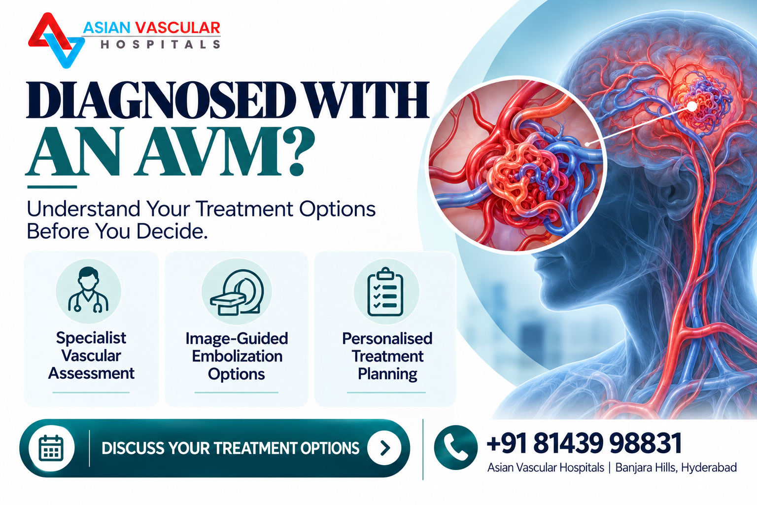
An arteriovenous malformation (AVM) is an abnormal connection between arteries and veins that can affect blood flow through the involved area. Depending on its location, size and structure, an AVM may cause swelling, pain, skin changes, bleeding or other complications. Some AVMs require active treatment, while others need specialist monitoring and an individualised management plan.
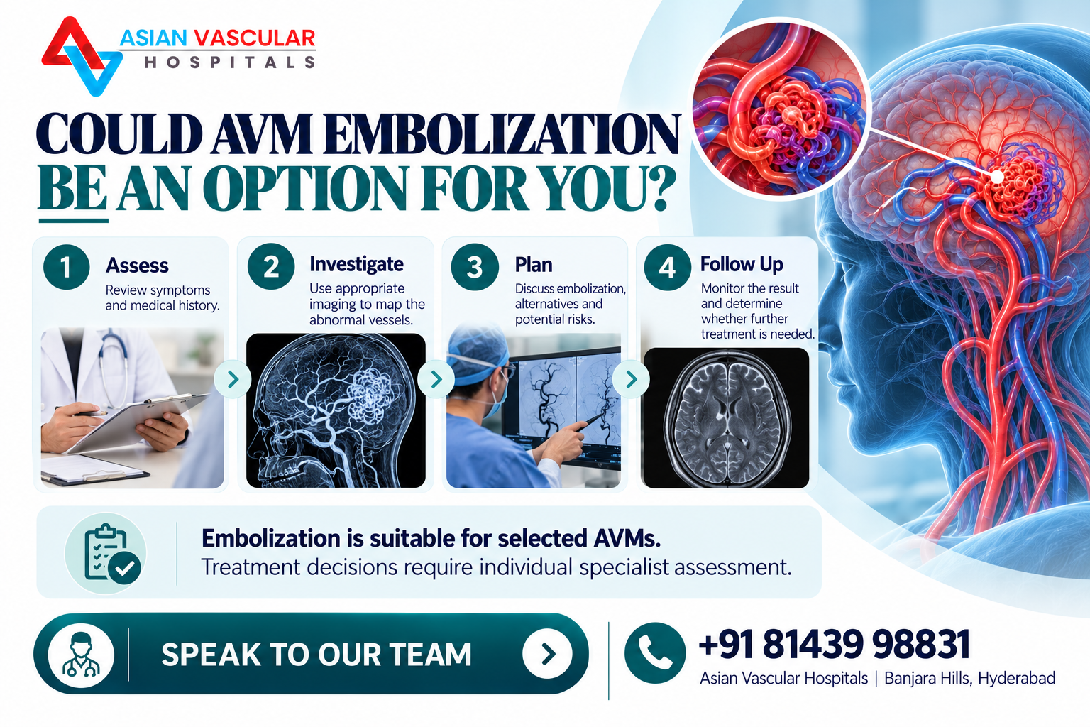
AVM embolization treatment is a minimally invasive technique that uses a catheter to deliver a material into selected abnormal blood vessels to reduce or block blood flow through the malformation. It may be used as a treatment on its own in selected cases or as part of a broader treatment plan.
However, embolization is not suitable for every AVM, and it does not always eliminate the malformation completely. The expected benefit, risks and possibility of additional procedures depend on the anatomy of the AVM and the specialist team’s assessment.
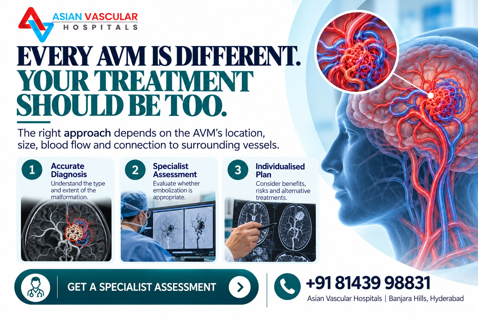
This guide explains how AVMs differ from other vascular malformations, how doctors assess them, what happens during an AVM embolization procedure, and what patients may expect during recovery.
What Is an Arteriovenous Malformation (AVM)?
An arteriovenous malformation is an abnormal network or connection of blood vessels in which blood passes from arteries into veins without the usual intervening capillary network.
Normally, arteries carry blood away from the heart, and capillaries allow oxygen and nutrients to pass into tissues before blood returns through veins. In an AVM, the abnormal connections can alter normal blood-flow patterns.
AVMs may occur in different parts of the body, including the limbs, skin, muscles and internal organs. Brain and spinal AVMs require specialised assessment because their location creates distinct risks and treatment considerations.
Depending on its location and characteristics, an AVM may cause:
- A visible or palpable mass
- Swelling or discomfort
- Skin discolouration or warmth
- Enlarged or prominent blood vessels
- Bleeding from an affected area
- Skin breakdown or ulceration in some cases
- Symptoms related to the affected organ
Some AVMs produce few noticeable symptoms, while others can cause significant problems. The absence of pain does not necessarily mean that an AVM is harmless.
How Is an AVM Different From Other Vascular Malformations?
Not all vascular malformations are AVMs, and different types may require different treatments.
The term vascular malformation describes a broader group of abnormalities involving blood vessels or lymphatic vessels. Correctly identifying the type of malformation is essential before treatment is planned.
| Type | What it involves | Treatment considerations |
|---|---|---|
| Arteriovenous malformation (AVM) | Abnormal connections between arteries and veins, often involving high-flow circulation | Embolization, surgery or combined treatment may be considered, depending on the anatomy and location |
| Venous malformation | Abnormally formed veins, commonly associated with slow blood flow | Observation, sclerotherapy or other selected treatments may be appropriate |
| Capillary malformation | Abnormal capillaries that may appear as flat red or purple skin patches | Laser treatment may be considered for selected lesions |
| Lymphatic malformation | Abnormal lymphatic channels or fluid-filled spaces | Management may include observation, sclerotherapy or other specialist procedures |
These conditions can sometimes look similar on the surface, but their blood-flow characteristics differ. For example, sclerotherapy is commonly used for selected venous malformations, whereas an AVM may require a different strategy because of its arterial connections and blood-flow pattern.
A clinical examination and appropriate imaging help establish the diagnosis and guide the treatment plan.
What Is AVM Embolization Treatment?
AVM embolization is a catheter-based procedure that delivers a specialised material into targeted blood vessels to reduce or stop blood flow through an abnormal vascular connection.
Rather than removing the malformation through an open operation, the specialist navigates a thin catheter through the blood vessels to reach the target area.
Depending on the AVM’s characteristics, embolization materials may include medical liquid agents, adhesives, particles, coils or other devices. The material selected depends on the type of malformation, its blood-flow pattern and the treatment objective.
Embolization may be considered to:
- Reduce blood flow through an AVM
- Manage selected symptoms or bleeding risks
- Treat certain malformations when embolization alone is appropriate
- Reduce blood flow before surgery in selected patients
- Form part of a combined treatment plan with surgery or radiosurgery when indicated
The goal is not identical for every patient. In some cases, embolization may achieve complete closure of the abnormal connection. In others, it may reduce blood flow without eliminating the entire malformation.
An important distinction: Embolization used for a peripheral AVM is not automatically the same treatment pathway used for a brain AVM. Cerebral AVMs require assessment by the appropriate cerebrovascular specialists, and treatment may involve an interventional neuroradiologist, neurosurgeon or other specialist team.
Who May Need AVM Embolization?
AVM embolization may be considered when a specialist determines that the potential benefits of reducing blood flow outweigh the procedural risks.
The decision may depend on:
- Location: Whether the AVM affects the skin, a limb, an internal organ, the brain or another area.
- Size and structure: The extent of the abnormal vascular network and its connections to surrounding vessels.
- Blood-flow characteristics: Whether the malformation has high-flow connections and how blood enters and exits it.
- Symptoms: Pain, swelling, bleeding, skin damage or other complications.
- Previous treatment: Whether the AVM has already been treated and whether it has persisted or returned.
- Treatment objectives: Whether embolization is intended as definitive treatment, symptom management or preparation for another procedure.
- Overall health: Other medical conditions and the individual’s ability to undergo the proposed procedure.
Not every AVM needs immediate intervention. For some patients, observation and periodic imaging may be appropriate. For others, the risks of leaving the malformation untreated may justify active management.
The decision should follow a specialist assessment rather than symptoms or appearance alone.
How Is an AVM Assessed Before Embolization?
Imaging is essential for understanding the AVM’s anatomy and deciding whether embolization is appropriate.
The exact assessment depends on the location of the malformation and the questions that need to be answered.
1. Clinical examination
The specialist may ask when the AVM was first noticed, whether it has changed in size, and whether symptoms such as pain, swelling, bleeding or skin changes have developed.
Examination may also assess visible vessels, skin temperature, swelling, tenderness and circulation in the affected area.
2. Doppler ultrasound
For selected superficial or limb vascular malformations, Doppler ultrasound can help assess blood-flow direction, velocity and the characteristics of the vessels.
It may provide useful information about whether a lesion has high-flow features, although ultrasound alone may not show the full extent of every AVM.
3. MRI or CT imaging
MRI or CT may be recommended to evaluate the extent of a malformation, its relationship to surrounding tissues and involvement of deeper structures.
The choice depends on the location, suspected diagnosis and the information needed for treatment planning.
4. Angiography
Catheter angiography may be used when detailed mapping of the abnormal blood vessels is needed, particularly for planning or performing embolization.
It helps the specialist understand the vessels supplying the AVM and the drainage pathways. The need for this investigation depends on the case.
Before proceeding, the team should explain the proposed procedure, expected benefits, alternatives and individual risks.
What Happens During the AVM Embolization Procedure?
The details of an AVM embolization procedure vary according to the location and complexity of the malformation. A typical catheter-based approach may involve the following stages.
Step 1: Preparation
The team reviews the imaging, medical history, medications, allergies and relevant blood-test results. Kidney function may need to be assessed if contrast material is planned.
The specialist and anaesthesia team explain whether local anaesthesia, sedation or general anaesthesia is appropriate.
Step 2: Accessing a blood vessel
A small puncture is usually made in a suitable blood vessel, commonly in the groin or arm, depending on the approach.
A catheter is introduced through this access point and guided through the vessels using imaging.
Step 3: Mapping the abnormal vessels
Contrast material may be injected to make the blood vessels visible on X-ray imaging. This helps the specialist identify the target vessels and assess how blood flows through the AVM.
Step 4: Delivering the embolic material
The catheter is carefully positioned, and the selected embolic material is delivered to the intended vessels.
The aim is to reduce or block abnormal blood flow while avoiding unintended blockage of healthy vessels. The specialist may use one or more treatment sessions, depending on the anatomy and treatment plan.
Step 5: Checking the result
Imaging is used to assess the immediate effect of treatment. The catheter is then removed, and the access site is managed to control bleeding.
The patient is monitored during recovery for pain, bleeding, changes in circulation and other possible complications.
The procedure does not always finish the entire course of treatment. Some AVMs require staged embolization, surgery or another treatment method, and further imaging may be necessary to assess the longer-term result.
What Are the Risks of AVM Embolization?
AVM embolization is a specialised procedure, and its risks vary according to the location, size and complexity of the malformation, the material used and the patient’s health.
Possible risks include:
- Bleeding or bruising: At the catheter insertion site.
- Blood-vessel injury: Damage to a vessel during catheter navigation or treatment.
- Unintended blockage of a healthy vessel: Embolic material may affect blood flow outside the intended target.
- Reduced blood supply to nearby tissue: This can lead to tissue injury or skin damage.
- Nerve injury: Depending on the location of the malformation and the surrounding anatomy.
- Infection: At the access site or, less commonly, in the treated area.
- Contrast-related complications: Allergic reactions or kidney-related concerns in susceptible patients.
- Incomplete treatment or recurrence: Some abnormal vessels may remain open or become apparent later.
- Further procedures: Additional embolization, surgery or another treatment may be needed.
For AVMs in the brain, specific risks can include stroke, bleeding within the brain and neurological injury. These risks require a separate, location-specific discussion with the appropriate cerebrovascular team.
This list is not exhaustive, and it does not mean that every patient will experience these complications. The treating specialist should explain which risks are most relevant to the individual case.
Recovery After Embolization: What Can Patients Expect?
Recovery after embolization depends on the AVM’s location, the extent of treatment, the anaesthesia used and whether additional procedures are required.
Some patients are discharged after a period of observation, while others need an overnight stay or longer monitoring.
Immediately after the procedure
The medical team checks the catheter insertion site, vital signs and circulation. Patients may be asked to keep the affected limb still for a period, depending on the access route and closure method.
Mild discomfort or bruising around the puncture site can occur. Temporary pain or swelling around the treated malformation may also develop.
During the first few days
The specialist may recommend rest, activity restrictions, prescribed pain relief and specific instructions for caring for the access site.
Patients should follow their discharge instructions rather than relying on a general recovery timeline. Activity restrictions differ depending on the procedure and individual circumstances.
Over the following weeks
Swelling, tenderness or skin changes may take time to settle. The treated AVM may not disappear immediately, and a reduction in symptoms does not necessarily confirm that the abnormal vessels have been completely closed.
Follow-up appointments help the team evaluate healing and decide whether further treatment or imaging is required.
There is no single recovery period that applies to every AVM embolization procedure. Ask the treating team when it is safe to return to work, exercise, driving and other usual activities.
When Should You Seek Medical Help After Embolization?
Follow the discharge instructions provided by the treating team. Seek urgent medical assessment if you experience:
- Bleeding that does not stop with the instructed first aid
- Rapidly increasing swelling or severe pain
- A newly cold, pale, blue or numb limb
- New weakness, difficulty speaking, changes in vision or other sudden neurological symptoms
- Fever with worsening redness, discharge or other signs of infection
- Any sudden or severe symptom that your medical team has warned you about
Sudden neurological symptoms, severe bleeding or signs of critically reduced circulation require emergency care. Do not wait for a routine follow-up appointment.
Is AVM Embolization a Permanent Treatment?
Embolization can completely close some AVMs, but in other cases it reduces blood flow without eliminating the entire malformation.
The likelihood of complete and durable treatment depends on factors such as the AVM’s anatomy, the location of abnormal connections, the treatment approach and whether additional procedures are needed.
A specialist may recommend follow-up imaging to determine how much of the malformation remains and whether further treatment is appropriate.
Even when an AVM appears successfully treated, follow-up may still be necessary. Patients should not assume that symptoms improving or the visible lesion shrinking means the AVM has been permanently eliminated.
Ask your specialist what the intended endpoint of treatment is, how it will be assessed and what follow-up schedule is recommended.
AVM Embolization vs Surgery: How Are They Different?
Embolization and surgery are different approaches, and neither is automatically the best choice for every AVM.
| Feature | Embolization | Surgery |
|---|---|---|
| Approach | A catheter is guided through blood vessels to deliver embolic material | A surgeon accesses and removes or treats the malformation directly |
| Main objective | Reduce or block abnormal blood flow | Remove or directly treat the abnormal vascular tissue when feasible |
| Possible role | Standalone treatment in selected cases or preparation for another procedure | May be considered when the AVM’s anatomy and location make surgery appropriate |
| Limitations | May not close the entire AVM and may require further treatment | Operative risks depend on the AVM’s location, extent and relationship to surrounding structures |
| Recovery | Depends on the procedure, access site and treatment extent | Depends on the operation and the area treated |
For some patients, a combination of treatments is recommended. Others may be better suited to monitoring or a different intervention.
The decision should be based on detailed imaging, the expected benefit, procedural risks and the patient’s priorities.
AVM Embolization Treatment at Asian Vascular Hospitals, Hyderabad
Asian Vascular Hospitals in Banjara Hills, Hyderabad, lists vascular malformations and embolization among its vascular care services.
However, the appropriate treatment depends on the specific type and location of the AVM. Before arranging a procedure, patients should confirm whether the hospital evaluates and treats their particular malformation and whether the required specialist team and facilities are available.
A consultation can help clarify the diagnosis, the investigations needed, available treatment options and whether referral to another specialist is appropriate.
What should you bring to your consultation?
If you have already undergone an assessment, bring:
- Previous Doppler ultrasound, MRI, CT or angiography reports
- Relevant scan images, not just the written report
- Details of previous procedures or treatments
- A current medication list
- Information about allergies and relevant medical conditions
- A list of symptoms and any recent changes
These details can help the clinical team understand your history and decide what further assessment may be needed.
Asian Vascular Hospitals
Banjara Hills, Hyderabad, Telangana
Appointments: +91 81439 98831
Explore the hospital’s vascular malformations treatment service for further information.
Frequently Asked Questions About AVM Embolization
- All Post
- blog
Related posts:
 Vascular Surgeon Vijayawada – Advanced Care by Dr. G.V. Praveen Kumar
 Early Stage Diabetic Foot Ulcer – Catch It Before It Gets Serious
 🧠 Peripheral Neuropathy Test – Detect Nerve Damage Early & Protect Your Health
 Laser Treatment for Varicose Veins Near Me – Asian Vascular Hospitals
 🌟 Best Treatment for Peripheral Neuropathy – Asian Vascular Hospitals
 Vascular Surgeon in Medchal: Advanced Care for Healthy Circulation
 Diabetic Foot Shoes: Comfort & Protection for Every Step
 👣 Podiatrist Hyderabad – Expert Foot & Ankle Care at Asian Vascular Hospitals