
Leg Swelling Isn’t Always a Muscle Problem. Sometimes It’s a Blood Clot.
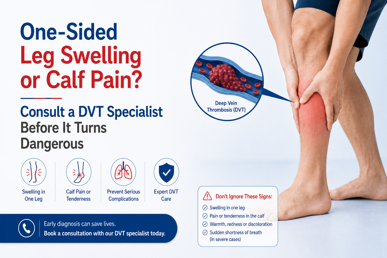
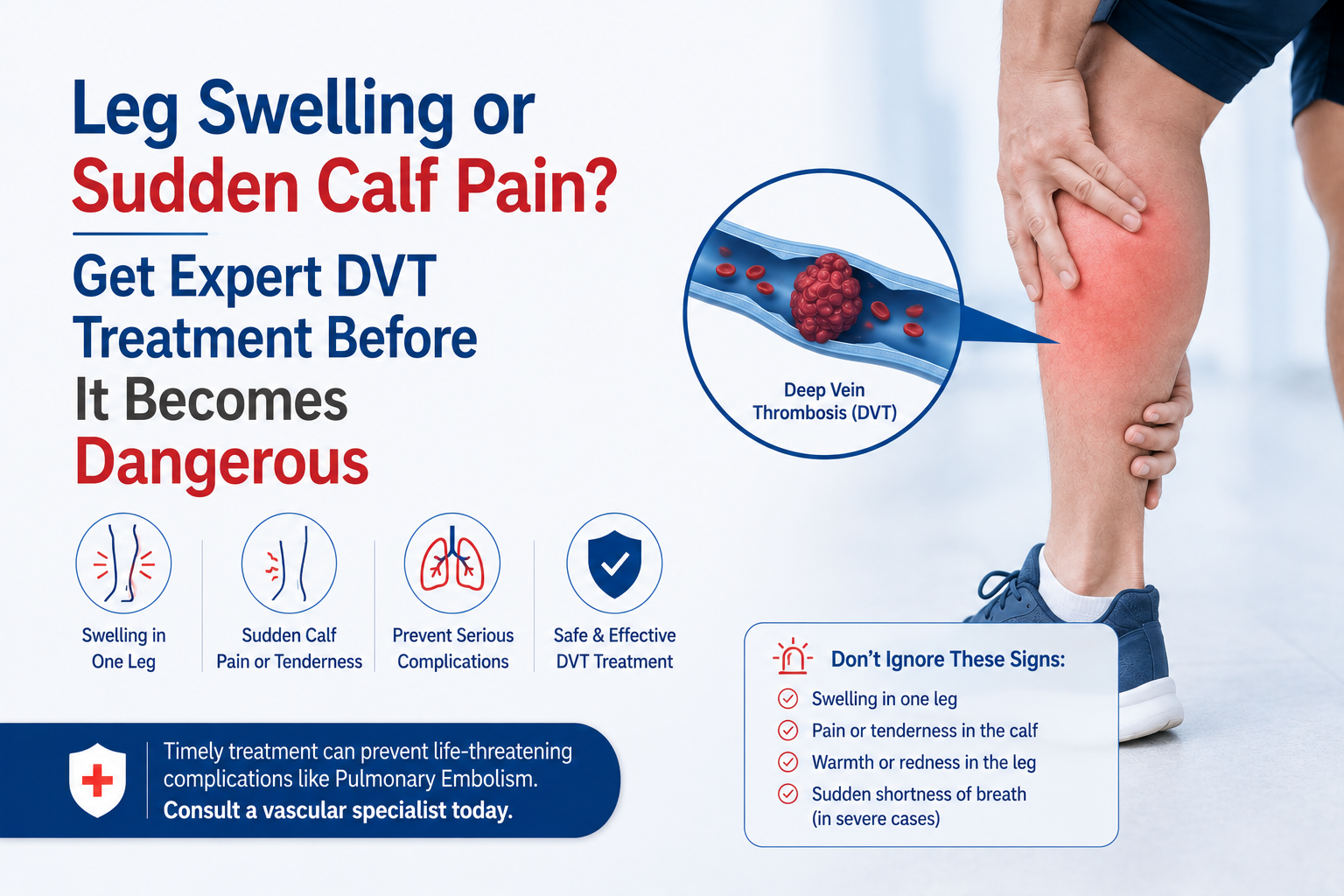
A swollen leg after a long journey, persistent calf pain, or unexplained heaviness can be easy to dismiss. However, these symptoms can sometimes point to Deep Vein Thrombosis (DVT)—a condition where a blood clot forms in a deep vein, usually in the leg.
The challenge is that DVT symptoms often overlap with muscle injuries, varicose veins, and circulation disorders. That’s why accurate dvt diagnosis is critical. The sooner a clot is identified, the sooner treatment can begin to prevent serious complications.
How Is DVT Diagnosed?
DVT diagnosis typically involves a clinical examination followed by imaging tests such as a doppler scan or ultrasound for dvt. In some cases, doctors may also recommend a blood clot test to assess the likelihood of clot formation before confirming the diagnosis with imaging.
Why Accurate DVT Diagnosis Matters
Not every swollen leg contains a blood clot.
At the same time, assuming symptoms are harmless can be dangerous.
A delayed dvt diagnosis may increase the risk of:
- Pulmonary embolism (clot reaching the lungs)
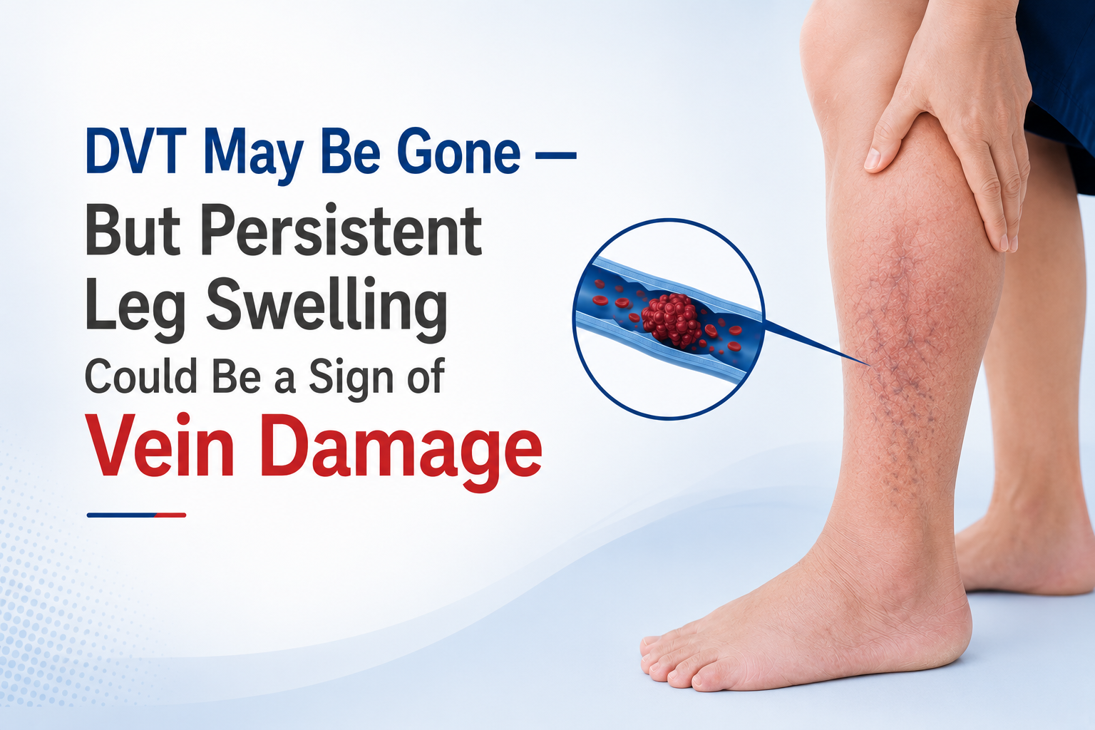
- Long-term vein damage
- Chronic leg swelling
- Post-thrombotic syndrome
- Emergency hospitalization
This is why vascular specialists focus on confirming the diagnosis quickly rather than guessing based on symptoms alone.
The First Step: Understanding Your Symptoms
Before any scan is performed, doctors assess the pattern of symptoms.
Signs that commonly trigger DVT evaluation:
- Swelling in one leg
- Persistent calf pain
- Warm skin over the affected area
- Redness or skin discoloration
- Sudden leg heaviness
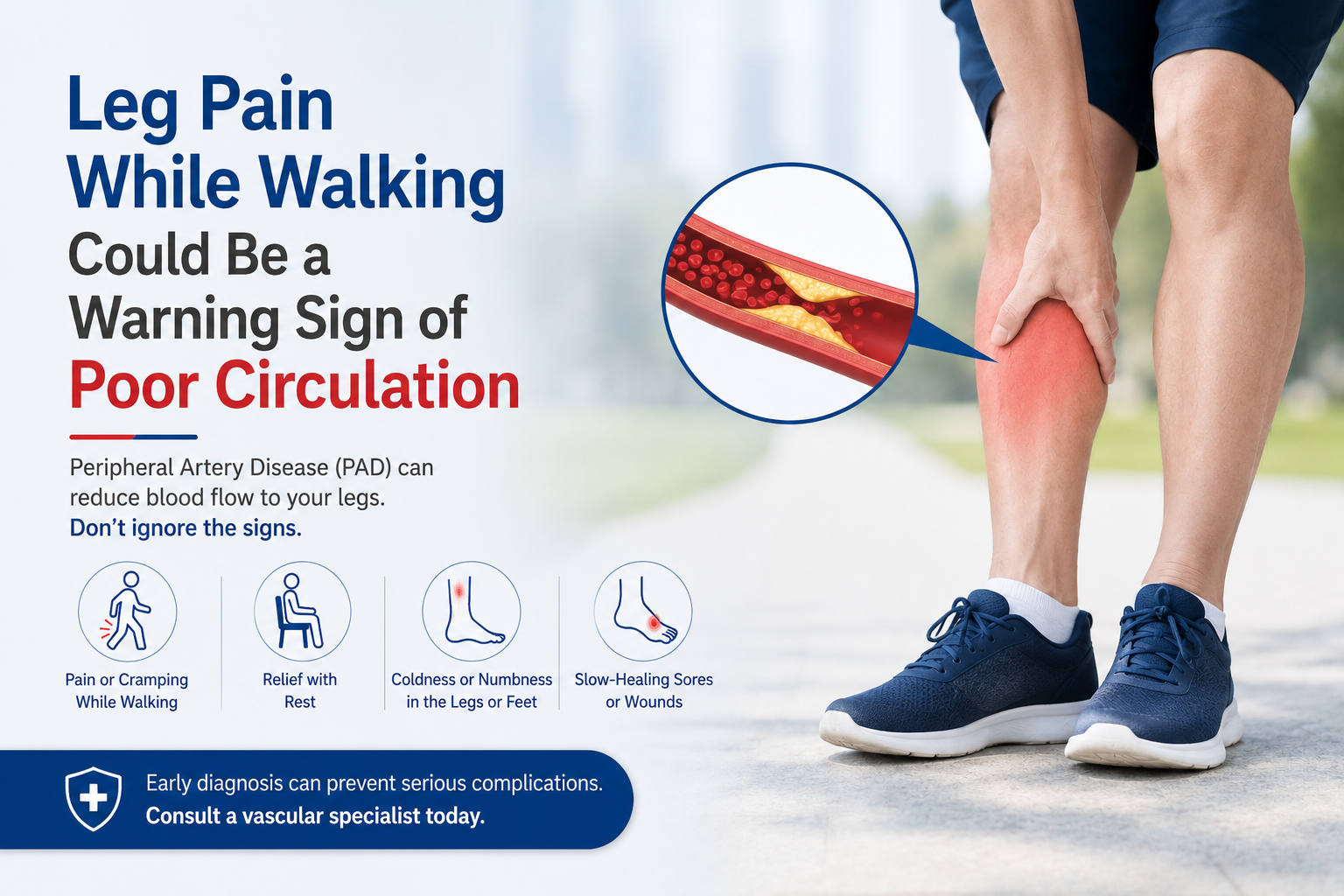
- Pain worsening while walking
These symptoms help determine whether urgent dvt diagnosis is required.
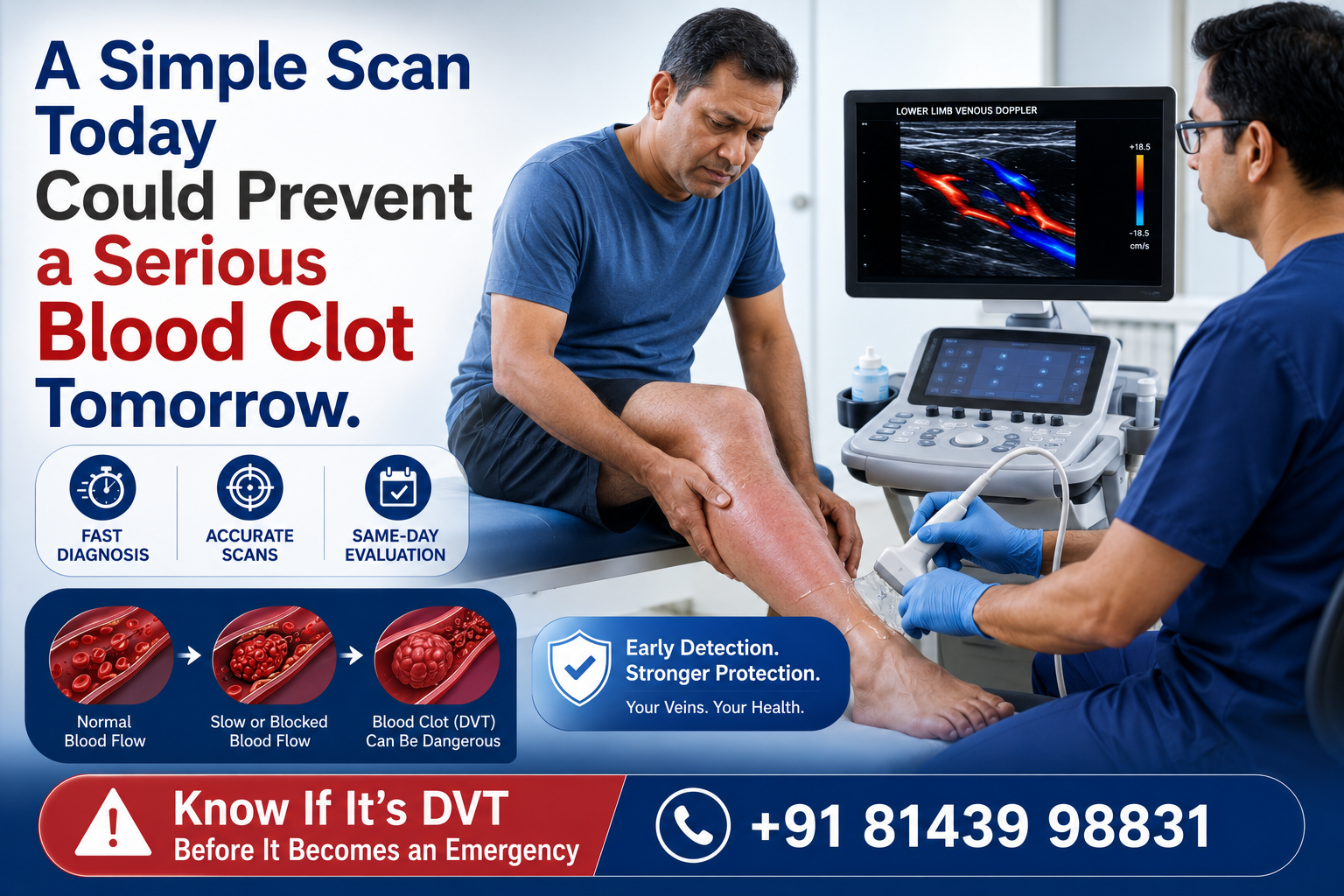
The Most Important Test: Doppler Ultrasound
Why Doppler Is Considered the Gold Standard
A doppler scan is usually the first and most important imaging test used for suspected DVT.
This test allows doctors to:
- Visualize blood flow inside veins
- Identify clot location
- Detect partial or complete blockages
- Assess circulation in real time
Because it is non-invasive, painless, and highly accurate, an ultrasound for dvt is often completed on the same day as consultation.
What Happens During an Ultrasound for DVT?
Many patients worry about the procedure, but it is simple and comfortable.
During the scan:
- Gel is applied to the skin
- A handheld probe is moved over the leg
- Blood flow is viewed on a monitor
- The specialist checks for clot-related blockage
The test usually takes less than 30 minutes and requires no recovery time.
Can a Blood Test Detect DVT?
A blood clot test known as the D-Dimer test may be recommended in selected cases.
What the test does:
- Measures clot-related substances in the blood
- Helps estimate clot probability
- Supports clinical decision-making
However, a blood clot test alone cannot confirm DVT.
Imaging is still required for accurate dvt diagnosis.
When Is CT Imaging Needed?
Most patients only require a doppler scan.
However, advanced imaging may be recommended when:
- The clot extends into the pelvis
- Symptoms are severe
- Pulmonary embolism is suspected
- Ultrasound findings are unclear
These scans help specialists create a complete treatment plan.
Common Myths About DVT Testing
“If the pain improves, the clot is gone.”
Not necessarily. Symptoms can fluctuate even when a clot remains present.
“A blood test is enough.”
A blood clot test helps assess risk but does not replace imaging.
“Only older people get DVT.”
DVT can occur at any age, especially after surgery, travel, pregnancy, or prolonged sitting.
What Happens After a DVT Diagnosis?
Once a clot is confirmed, treatment begins based on:
- Clot size
- Vein involvement
- Risk factors
- Overall health condition
Treatment may include:
- Blood thinners
- Compression therapy
- Catheter-based clot removal
- Advanced vascular interventions
Early treatment significantly improves outcomes.
Real Clinical Insight
One of the most common scenarios seen in vascular practice is a patient treating calf pain as a muscle strain for several days before seeking help.
In many cases, a simple ultrasound for dvt reveals a clot that could have progressed if left untreated. Early imaging often makes the difference between routine treatment and emergency intervention.
When Should You Request a Doppler Scan?
Consider immediate evaluation if you have:
✔ One-sided leg swelling
✔ Calf pain without injury
✔ Warm or red skin
✔ Recent surgery or hospitalization
✔ Long-distance travel history
✔ Previous DVT
Prompt dvt diagnosis allows specialists to intervene before complications develop.
Why Choose a Dedicated Vascular Hospital for DVT Diagnosis?
At Asian Vascular Hospitals, DVT evaluation is performed using advanced vascular imaging and specialist-led assessment.
Patients benefit from:
✔ Same-day vascular consultation
✔ Advanced doppler scan facilities
✔ Accurate ultrasound for dvt
✔ Emergency clot evaluation
✔ Personalized treatment planning
✔ 24/7 vascular care support
📞 Get Expert DVT Evaluation Before Complications Develop
If you are experiencing unexplained leg swelling, calf pain, or symptoms suggestive of a blood clot, don’t rely on guesswork.
Led by Dr. G. V. Praveen Kumar, Asian Vascular Hospitals provides advanced dvt diagnosis, vascular imaging, and minimally invasive clot treatment under one roof.
📞 Call Now: +91 81439 98831
🌐 Visit: asianvascularhospitals.com
🚑 24/7 Emergency Vascular Care Available
- All Post
- blog

Related posts:
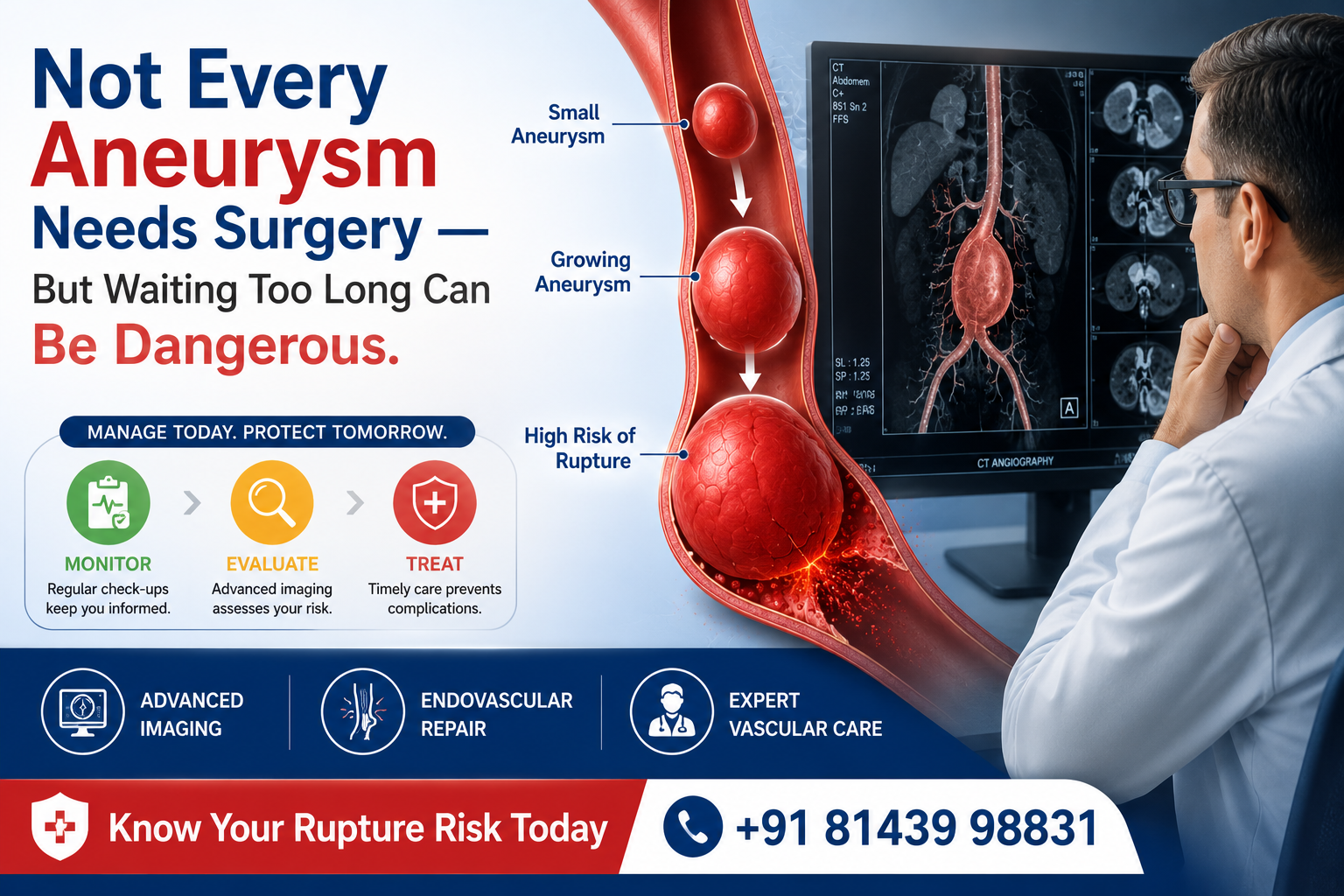
 Aneurysm Treatment in Hyderabad: Expert Care for Your Vascular Health
 Best Vascular Surgeon – Everything You Need to Know Before Choosing One
 Varicose Veins Treatment Cost – Affordable Laser Surgery in Hyderabad
 Peripheral Neuropathy: Causes, Symptoms, and Treatment Options
 🦶 Diabetic Foot Care at Asian Vascular Hospitals, Hyderabad
 Vascular Surgeon in Karimnagar: Advanced Vascular Care for Healthier Living
 Vascular Surgeon in Bhuvanagiri: Advanced Vein & Artery Care
 Varicose Veins Medical Treatment – Advanced Options for Lasting Relief
